Understanding Extrarenal Pelvis: A Common Kidney Variant
Receiving medical scan results can often feel like deciphering a foreign language, filled with complex terms that leave you more confused than informed. One such term you might encounter, particularly if you've had a CT scan or ultrasound of your kidneys, is "extrarenal pelvis." This phrase, while sounding potentially alarming, often refers to a perfectly normal anatomical variation. Understanding what an extrarenal pelvis is, why it occurs, and what it means for your health is crucial to alleviate any unnecessary worry and empower you with knowledge about your body.
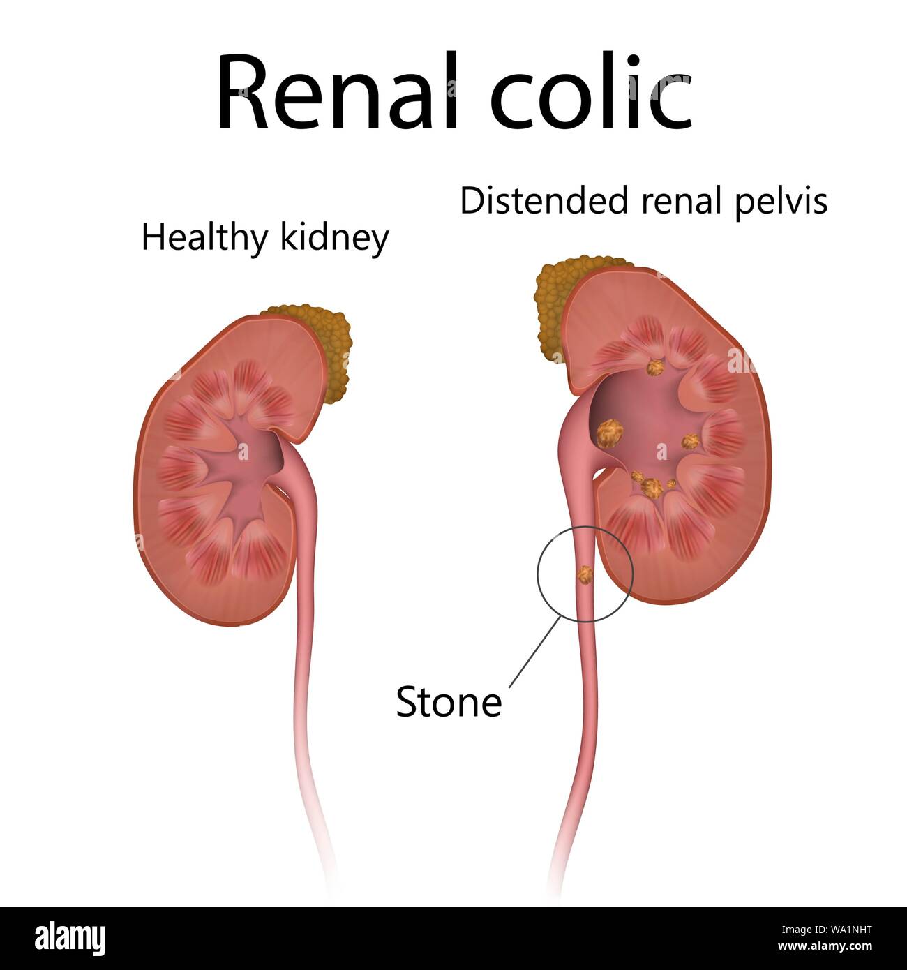
The kidneys are vital organs, diligently filtering waste from your blood and producing urine. Central to this process is the renal pelvis, a funnel-shaped structure within the kidney that collects urine before it flows into the ureter and down to the bladder. In some individuals, however, this collecting area extends beyond the confines of the kidney itself, leading to what is termed an extrarenal pelvis. This article aims to demystify this common finding, providing clear, comprehensive information based on typical medical understanding and diagnostic interpretations.
The Kidneys and Their Pelvis: A Brief Overview
To truly grasp the concept of an extrarenal pelvis, it helps to first understand the basic anatomy and function of your kidneys. Located on either side of your spine, just below the rib cage, these bean-shaped organs play a critical role in maintaining your body's internal balance. They filter about 120 to 150 quarts of blood daily, removing waste products and excess water to produce urine. This urine then travels down through narrow tubes called ureters to the bladder, where it is stored until urination.
Within each kidney, the filtering units, known as nephrons, produce urine. This urine then drains into a central collecting area known as the renal pelvis. Think of the renal pelvis as the "middle where everything centers" within the kidney, as it collects all the urine made by filtering blood. From this central point, the urine is then channeled out through the ureters to the bladder. The shape and position of this renal pelvis are generally consistent, but like many biological structures, variations can occur. One such variation is the extrarenal pelvis, where this collecting structure extends outside the kidney's main body, making it more prominent or "bigger size than normal" in appearance on imaging studies.
What Exactly Is an Extrarenal Pelvis?
An extrarenal pelvis is essentially an anatomical variation where the renal pelvis, instead of being entirely contained within the kidney's substance, protrudes or extends outwards from the kidney's hilum (the indented area where blood vessels and nerves enter and exit). This means that a portion of the collecting system is located outside the kidney's fibrous capsule. It's not a disease or a pathology in itself, but rather a different way the kidney has developed. As noted in various scan impressions, an "extrarenal type of renal pelvices is considered which is a variant of normal advice."
This variation is quite common and is often discovered incidentally during imaging studies like a CT scan or ultrasound performed for other reasons, such as investigating "pelvis pain" or as part of a routine check-up. For instance, it might be mentioned on a "CT study" or even observed during a "33 week baby ultrasound shows" as a developmental finding. The key takeaway here is that an extrarenal pelvis is, in most cases, a "very common variation of renal development" and is considered a normal anatomical variant.
Diagnosing an Extrarenal Pelvis and Its Implications
The diagnosis of an extrarenal pelvis typically comes from medical imaging. When you have a CT scan, ultrasound, or MRI of your abdomen or kidneys, radiologists look at the structure and position of your organs. If the renal pelvis is seen to extend beyond the kidney's usual boundaries, it will be noted in the scan report. Phrases like "The following is on my scan report what does it mean" often lead to explanations from healthcare providers, who will clarify that "an enlarged extrarenal pelvis is a normal variant."
However, while generally a normal variant, it's important to understand that its presence can sometimes be associated with other conditions, or it might be misinterpreted as a problem. For example, the larger, more prominent appearance of an extrarenal pelvis can sometimes be confused with hydronephrosis, a swelling of the kidney due to urine backup. This is why careful interpretation by a radiologist and a clinician is essential. The distinction between a normal extrarenal pelvis and actual hydronephrosis is critical for appropriate management.
Bilateral Extrarenal Pelvis: What It Means
It's not uncommon for an extrarenal pelvis to be present in both kidneys, a condition referred to as a "bilateral extrarenal pelvis." When a member asks, "What is a bilateral extrarenal pelvis?", it simply means that this anatomical variation exists on both the right and left sides. Just like a unilateral (one-sided) extrarenal pelvis, a bilateral presentation is also typically considered a normal variant and does not inherently signify a problem. The functionality of both kidneys is usually unaffected by this anatomical difference.
However, as with any anatomical variation, healthcare providers will assess the overall kidney health and function. If other symptoms or findings are present, they might investigate further. The key is that the bilateral nature itself is not a cause for alarm, but rather a characteristic of the individual's unique renal anatomy.
Extrarenal Pelvis and UPJ Obstruction: Understanding the Link
While an extrarenal pelvis is usually benign, it's crucial to acknowledge that "an enlarged extrarenal pelvis is a normal variant but can be associated sometimes with a UPJ obstruction." The ureteropelvic junction (UPJ) is the area where the renal pelvis narrows and connects to the ureter. An obstruction at this point can hinder the flow of urine, leading to a buildup of urine in the kidney (hydronephrosis). The presence of an extrarenal pelvis, due to its more external position, might theoretically make it more susceptible to kinking or compression, or it might simply be present alongside a true UPJ obstruction.
This is why, if there are symptoms like pain, recurrent infections, or if the imaging shows signs of urine stasis, further evaluation is warranted. A "renal scan may help evaluate for an obstructive process," providing dynamic information about how urine flows through the kidney and out to the bladder. This scan can differentiate between a non-obstructive extrarenal pelvis and one that is genuinely causing a blockage.
Symptoms and Concerns: When to Seek Medical Advice
In the vast majority of cases, an extrarenal pelvis causes no symptoms whatsoever. It's an incidental finding that doesn't impact kidney function or overall health. Many people live their entire lives unaware they have this anatomical variation. However, if you experience "pain associated with extra renal pelvis," or other concerning symptoms, it's vital to consult a healthcare professional. The pain you're experiencing might be due to an unrelated issue, or in rare cases, it could be linked to complications that are more likely to occur with an extrarenal pelvis.
Potential symptoms that warrant medical attention include:
- Flank pain (pain in the side or back, just below the ribs)
- Recurrent urinary tract infections (UTIs)
- Blood in the urine (hematuria)
- Fever and chills, especially if accompanied by flank pain
- Nausea and vomiting
These symptoms could indicate complications such as a kidney stone or an actual obstruction, which might be more pronounced or symptomatic in the presence of an extrarenal pelvis. For example, if a "mid pole calculus is noted in my right kidney measuring 8.7 mm and mild hydronephrosis with extra renal pelvis is seen in the both kidneys," the pain is likely due to the kidney stone and the associated hydronephrosis, rather than the extrarenal pelvis itself being the primary cause of discomfort. The extrarenal pelvis simply provides the anatomical context.
Managing an Extrarenal Pelvis and Associated Conditions
If an extrarenal pelvis is found incidentally and you have no symptoms, no specific treatment or management is typically required. It's simply noted as part of your medical record. However, if there are associated findings or symptoms, the management will focus on those specific issues.
For instance, if a kidney stone is present, treatment will focus on managing the stone, which might involve pain medication, watchful waiting for the stone to pass, or interventions like lithotripsy or surgery. If mild hydronephrosis is noted alongside an extrarenal pelvis, especially if asymptomatic, it might be monitored periodically with follow-up ultrasounds to ensure it doesn't progress or become obstructive. The key is to differentiate between a benign anatomical variant and a pathological condition that requires intervention.
The Role of Further Investigation
When there's a question about whether an extrarenal pelvis is truly benign or if it's contributing to symptoms or obstruction, further investigations might be recommended. As mentioned, a "renal scan may help evaluate for an obstructive process." This type of scan, often a diuretic renogram, involves injecting a small amount of radioactive tracer and then a diuretic to see how quickly the tracer (and thus urine) clears from the kidney. This helps doctors determine if there's a functional obstruction or just a dilated but non-obstructive collecting system.
Other investigations might include repeat imaging, urine tests to check for infection, or blood tests to assess kidney function. The decision for further tests is always made by a healthcare professional based on the individual's symptoms, medical history, and initial imaging findings. It's about ensuring that the extrarenal pelvis isn't masking or contributing to a more significant underlying issue.
Understanding Your Scan Report and Doctor's Explanation
It's common to feel overwhelmed by medical reports. Phrases like "I got an ultrasound earlier today and got results back, but doctor didn't explain everything" highlight the need for clear communication. When your report mentions an extrarenal pelvis, your doctor should explain that it's a "normal variant" and what that implies for your health. Don't hesitate to ask questions if you don't understand something in your report or your doctor's explanation. For instance, if your report states, "The right renal pelvis is likely duplicated with mild anterior malrotation of the lower," this indicates another type of anatomical variation that your doctor should clarify in detail.
A good doctor will take the time to address your concerns, explain the findings in layman's terms, and outline any necessary follow-up. As "a doctor has provided 1 answer a member" or "a doctor has provided 1 answer a" indicates, healthcare professionals are there to interpret these findings for you and guide you on the best course of action.
Living with an Extrarenal Pelvis
For most individuals, living with an extrarenal pelvis means living without any special considerations or lifestyle changes. It's a part of your unique anatomy, much like having a specific eye color or height. You don't need to restrict your diet, activities, or take any particular medications because of it. Regular check-ups and maintaining general kidney health through adequate hydration and a balanced diet are always good practices for everyone, regardless of whether they have an extrarenal pelvis.
The main significance of knowing you have an extrarenal pelvis is for future medical contexts. If you ever develop kidney-related symptoms, your healthcare providers will be aware of this anatomical variation, which can help them in their diagnostic process. It ensures that a normal variant isn't mistakenly identified as a problem, and conversely, that any actual problems aren't overlooked because of the variant.
Key Takeaways About Extrarenal Pelvis
In summary, the journey of understanding medical terms like "extrarenal pelvis" begins with clear, reliable information. Here are the essential points to remember:
- The renal pelvis is the kidney's central collecting area for urine, which then flows to the bladder via the ureters.
- An extrarenal pelvis is an anatomical variation where a portion of the renal pelvis extends outside the kidney's main body.
- It is a common finding, often detected incidentally during imaging studies like CT scans or ultrasounds.
- In most cases, an extrarenal pelvis is a normal variant and does not cause any symptoms or require treatment.
- While generally benign, its presence can sometimes be associated with conditions like UPJ obstruction or kidney stones, especially if symptoms like pain or recurrent infections are present.
- If symptoms occur, further evaluation such as a renal scan may be recommended to differentiate between a normal variant and an obstructive process.
- Understanding your scan report and discussing findings with your doctor is crucial for peace of mind and appropriate management.
Ultimately, an extrarenal pelvis is usually a benign anatomical quirk. It's a testament to the diverse ways human bodies can develop. While the initial mention in a scan report might cause concern, armed with accurate information, you can approach your health with confidence and clarity, knowing that this common variation is, for most, just another unique aspect of their perfectly functional body.
Conclusion
Navigating medical terminology can be daunting, but understanding conditions like an extrarenal pelvis is key to managing your health effectively. We've explored how this common anatomical variation, where the kidney's urine-collecting basin extends beyond its usual confines, is typically a normal finding rather than a cause for alarm. From its detection on routine scans to its potential, albeit rare, association with issues like UPJ obstruction or kidney stones, the overarching message remains: an extrarenal pelvis is, for the vast majority, simply a unique aspect of their renal anatomy that functions perfectly well.
Remember, while this article provides comprehensive information, it is not a substitute for professional medical advice. Always discuss your specific scan results and any symptoms with your healthcare provider. Their expertise is invaluable in interpreting your individual situation and guiding your care. If you found this article helpful in demystifying "extrarenal pelvis," please consider sharing it with others who might benefit from this knowledge. We also invite you to leave a comment below with your thoughts or questions, or explore other health-related articles on our site for more insights into common medical conditions.


Detail Author:
- Name : Mr. Joan Zieme
- Username : halie68
- Email : novella50@will.info
- Birthdate : 2007-04-21
- Address : 3641 Joaquin Crossing Suite 624 Leraburgh, RI 64791-2651
- Phone : 731-794-4626
- Company : Brekke Ltd
- Job : Production Control Manager
- Bio : Aperiam laudantium qui ea. Assumenda temporibus maiores nostrum quidem quam laborum. Enim porro laudantium est corrupti explicabo.
Socials
twitter:
- url : https://twitter.com/kelly_ebert
- username : kelly_ebert
- bio : Libero sed magnam dolorum eos nam. Distinctio id dolores est est quibusdam et.
- followers : 5274
- following : 1123
instagram:
- url : https://instagram.com/kelly.ebert
- username : kelly.ebert
- bio : In aut eveniet fugiat qui cumque voluptas. Amet omnis illum tempora dignissimos.
- followers : 5750
- following : 2443